
Histology of the eye
 Figure 1: Tapetum fibrosum of the canine eye. 1: Sclera composed of white dense fibrous tissue 2: Lamina fusca 3: Choroid with capillaries 4: Tapetum cellulosum 5: Photosensitive layers of the retinal layers 6: Pigment layer 7: Rods and cones 8: Nuclei of rods and cones 9: Outer synaptic layer 10: Bipolar nerve cell nuclei 11: Inner synaptic layer 12: Optic nerve cells 13: Optic nerve fibres. H&E x160
Source: Aughey E. and Frye E.L. (2001) Comparative veterinary histology, 1st edn., London: Manson Publishing Ltd.
Figure 1: Tapetum fibrosum of the canine eye. 1: Sclera composed of white dense fibrous tissue 2: Lamina fusca 3: Choroid with capillaries 4: Tapetum cellulosum 5: Photosensitive layers of the retinal layers 6: Pigment layer 7: Rods and cones 8: Nuclei of rods and cones 9: Outer synaptic layer 10: Bipolar nerve cell nuclei 11: Inner synaptic layer 12: Optic nerve cells 13: Optic nerve fibres. H&E x160
Source: Aughey E. and Frye E.L. (2001) Comparative veterinary histology, 1st edn., London: Manson Publishing Ltd.
 Figure 2: Tapetum fibrosum of the eye in a horse. 1: Pigment layer of the retina 2: compact layer of fibrous connective tissue 3: Choroid with blood vessels and some pigment cells. H&E. x100
Source: Aughey E. and Frye E.L. (2001) Comparative veterinary histology, 1st edn., London: Manson Publishing Ltd.
 Figure 3: Posterior surface of a canine cornea. 1: Substantia propria 2: Basement membrane (Descemet's) 3: Simple squamous endothelium. H&E. x62.5
Source: Aughey E. and Frye E.L. (2001) Comparative veterinary histology, 1st edn., London: Manson Publishing Ltd.
 Figure 4: Lens of a canine. 1: Anterior epithelium 2: Nuclei of lens fibres. H&E. x62.5
Source: Aughey E. and Frye E.L. (2001) Comparative veterinary histology, 1st edn., London: Manson Publishing Ltd.
 Figure 5: Ciliary processes of canine. 1: Long ciliary processes extend from the ciliary body. The arrow is pointing to the epithelium 2: Ciliary muscle. H&E. x62.5
Source: Aughey E. and Frye E.L. (2001) Comparative veterinary histology, 1st edn., London: Manson Publishing Ltd.
 Figure 6: Canine iris. 1: Anterior surface is covered by flattened fibrocytes 2: Core of iris is vasculature connective tissue. 3: Posterior surface epithelium is two layers of cells and part of retina. There is presence of pigments. H&E. x62.5
Source: Aughey E. and Frye E.L. (2001) Comparative veterinary histology, 1st edn., London: Manson Publishing Ltd.
 Figure 7: Canine iris. Pigment in this picture is different from the pigment in figure 6.
Source: Aughey E. and Frye E.L. (2001) Comparative veterinary histology, 1st edn., London: Manson Publishing Ltd.
 Figure 8: Optic disc of a horse. 1: Scleral connective tissue 2: Optic nerve bundles. H&E. x25
Source: Aughey E. and Frye E.L. (2001) Comparative veterinary histology, 1st edn., London: Manson Publishing Ltd.

Source: Maggs D.J., Miller P.E. and Ofri R. (2013) Slatter's fundamentals of veterinary ophthalmology, 5th edn., Missouri: Elsevier.
 Figure 10: Fovea centralis of a monkey. FC: Fovea centralis; C: cones; Ch: Choroid; 1: Pigmented epithelium 2: Lamina of cones 3: External limiting membrane 4: Outer nuclear layer 5: Outer plexiform layer 8: Ganglion cell layers 10: Inner limiting membrane. x132
Source: Gartner L.P. and Hiatt J.L. (2009) Color atlas of histology, 5th edn., Philadelphia: Lippincott William & Wilkins.
 Figure 11: Optic nerve. S: Sclera ON: Optic nerve U: Uveal tract D: Dura mater P: Pia-arachnoid A: central artery of retina.
Source: Young B. and Deaken P.J. (2002) Wheater's functional histology: A text and colour atlas, 5th edn., Edinburgh: Churchill Livingstone Elsevier .
Figure 11: Optic nerve. S: Sclera ON: Optic nerve U: Uveal tract D: Dura mater P: Pia-arachnoid A: central artery of retina.
Source: Young B. and Deaken P.J. (2002) Wheater's functional histology: A text and colour atlas, 5th edn., Edinburgh: Churchill Livingstone Elsevier .
 Figure 12: Lacrimal gland separated into lobes and lobules (LO) by connective tissue (CO). SA: serous acini N: nuclei. x132
Source: Gartner L.P. and Hiatt J.L. (2009) Color atlas of histology, 5th edn., Philadelphia: Lippincott William & Wilkins.
Figure 12: Lacrimal gland separated into lobes and lobules (LO) by connective tissue (CO). SA: serous acini N: nuclei. x132
Source: Gartner L.P. and Hiatt J.L. (2009) Color atlas of histology, 5th edn., Philadelphia: Lippincott William & Wilkins.

 Figure 14: Avian eye. 1: Choroid 2: Retinal layers 3: Pigment layer 4: Rods and cones 5: Nuclei of the rods and cones 6: Outer synaptic layer 7: bipolar nerve 8: Inner synaptic layer 9: Optic nerve cells 10: Optic nerve fibres H&E. x125
Source: Aughey E. and Frye E.L. (2001) Comparative veterinary histology, 1st edn., London: Manson Publishing Ltd.
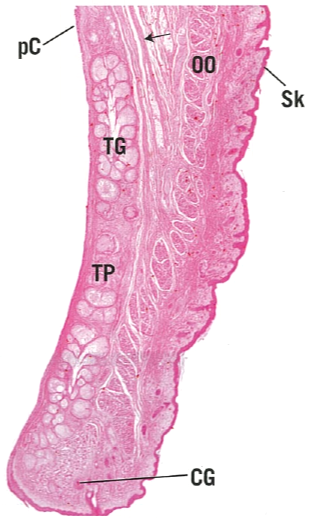 Figure 15: Eyelid. PC: Palpebral conjunctiva; OO: Orbicularis oculi; SK: Skin; TG: ; TP: ; CG: . x14
Source: Gartner L.P. and Hiatt J.L. (2009) Color atlas of histology, 5th edn., Philadelphia: Lippincott William & Wilkins.
 Figure 16: Eyelid of a horse. 1: Stratified columnar epithelium with mucus secreting cells of conjunctiva 2: Sebaceous glands in the lamina propria 3: Tarsal plate. H&E. x25.
Source: Aughey E. and Frye E.L. (2001) Comparative veterinary histology, 1st edn., London: Manson Publishing Ltd.
Figure 16: Eyelid of a horse. 1: Stratified columnar epithelium with mucus secreting cells of conjunctiva 2: Sebaceous glands in the lamina propria 3: Tarsal plate. H&E. x25.
Source: Aughey E. and Frye E.L. (2001) Comparative veterinary histology, 1st edn., London: Manson Publishing Ltd.
 Figure 17: Nictitating membrane (third eyelid) of a canine. 1: Hyaline cartilage 2: Seromucus-secreting glands in the lamina propria. H&E. x50
Source: Aughey E. and Frye E.L. (2001) Comparative veterinary histology, 1st edn., London: Manson Publishing Ltd.
Figure 17: Nictitating membrane (third eyelid) of a canine. 1: Hyaline cartilage 2: Seromucus-secreting glands in the lamina propria. H&E. x50
Source: Aughey E. and Frye E.L. (2001) Comparative veterinary histology, 1st edn., London: Manson Publishing Ltd.
 Figure 18: Nictitating membrane (third eyelid) of a horse. 1: Anterior conjunctiva surface. 2: Elastic cartilage plate. 3: Posterior conjunctival surface. 4: Lamina propria. H&E. x7.5
Source: Aughey E. and Frye E.L. (2001) Comparative veterinary histology, 1st edn., London: Manson Publishing Ltd.
References
Source: Aughey E. and Frye E.L. (2001) Comparative veterinary histology, 1st edn., London: Manson Publishing Ltd.
Source: Aughey E. and Frye E.L. (2001) Comparative veterinary histology, 1st edn., London: Manson Publishing Ltd.
Source: Aughey E. and Frye E.L. (2001) Comparative veterinary histology, 1st edn., London: Manson Publishing Ltd.
Source: Aughey E. and Frye E.L. (2001) Comparative veterinary histology, 1st edn., London: Manson Publishing Ltd.
Source: Aughey E. and Frye E.L. (2001) Comparative veterinary histology, 1st edn., London: Manson Publishing Ltd.
Source: Aughey E. and Frye E.L. (2001) Comparative veterinary histology, 1st edn., London: Manson Publishing Ltd.
Source: Aughey E. and Frye E.L. (2001) Comparative veterinary histology, 1st edn., London: Manson Publishing Ltd.
Figure 9: Layers of the retina.
Source: Gartner L.P. and Hiatt J.L. (2009) Color atlas of histology, 5th edn., Philadelphia: Lippincott William & Wilkins.
Figure 13: Avian eye. 1: Sclera with hyaline cartilage. 2: Choroid. H. & E. x62.5.
Source: Aughey E. and Frye E.L. (2001) Comparative veterinary histology, 1st edn., London: Manson Publishing Ltd.
Source: Aughey E. and Frye E.L. (2001) Comparative veterinary histology, 1st edn., London: Manson Publishing Ltd.
Source: Gartner L.P. and Hiatt J.L. (2009) Color atlas of histology, 5th edn., Philadelphia: Lippincott William & Wilkins.
Source: Aughey E. and Frye E.L. (2001) Comparative veterinary histology, 1st edn., London: Manson Publishing Ltd.
References
- Aughey E. and Frye E.L. (2001) Comparative veterinary histology, 1st edn., London: Manson Publishing Ltd.
- Gartner L.P. and Hiatt J.L. (2009) Color atlas of histology, 5th edn., Philadelphia: Lippincott William & Wilkins.
- Maggs D.J., Miller P.E. and Ofri R. (2013) Slatter's fundamentals of veterinary ophthalmology, 5th edn., Missouri: Elsevier.
- Young B. and Deaken P.J. (2002) Wheater's functional histology: A text and colour atlas, 5th edn., Edinburgh: Churchill Livingstone Elsevier .